|
Clinical endoscope for two-photon image-guided femtosecond laser surgery |
 |
|
Current Research
|
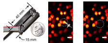
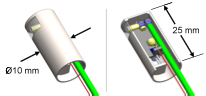
Left: Photograph of the first probe prototype, measuring just 10 × 15 × 40 mm3. Center: A two-photon fluorescence image of cancer cells taken with the prototype, indicating a cell targeted for ablation. Right: The same region, after delivering one high-energy surgery pulse to the targeted cell. The targeted cell was instantly destroyed while neighboring cells were left intact. Below: A preliminary model of a clinically packaged endoscope for treatment of vocal fold scars.
|
Two-photon microscopy is emerging as a useful imaging technique for diagnostic imaging of biological tissues. Additionally, femtosecond laser pulses have proven to be much more efficient in ablating tissue for microsurgery, thus greatly reducing collateral damage. Thus far, integration of these techniques into clinically-useful tools has been extremely limited. The aim of this project is to develop small flexible probes capable of delivering ultrashort laser pulses into the body for combined imaging and microsurgery. Currently, we are focused on two key clinical applications: treatment of small cancerous lesions and treatment of vocal fold scarring. This project has strong optical design and experimental components. The nature of the work is very multidisciplinary and includes fields such as MEMS devices, photonic crystal structures, manufacturing, signal processing, and cell biology. There are currently several proposals being reviewed to provide funding for this project. |
|