|
Nerve regeneration studies in Caenorhabditis elegans using laser axotomy and RNA interference |
 |
|
Current Research
|
|
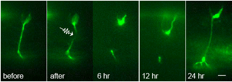 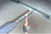
Top: Nerve regeneration after femtosecond laser axotomy in live C. elegans. Fluorescence images of GFP labeled axons before, after, and following axotomy. Bottom: Rendition of a trapped worm undergoing axotomy in a microfluidic chip.
|
In the United States, nearly 5 million people suffer from neurodegenerative diseases and another half?million have suffered from some type of nerve injury. To this day, the mechanisms underlying nerve regeneration and degeneration remain largely unknown. Searching for the genes involved in these mechanisms will help us to better understand the causes of these debilitating diseases and thus pave the way for successful treatments and preventions. The best conditions for studying nerve regeneration would be met if one could sever axons or dendrites in a controlled manner and study what genes or molecules affect their regrowth in vivo in a simple organism. The soil nematode Caenorhabditis elegans (C. elegans) is an ideal model organism for such study. They are transparent to visible light; their nervous system comprises only 302 neurons and is entirely mapped; and their genetic versatility allows for gene manipulations. In our animal model, C. elegans, we are investigating mutant worms and using RNA interference, along with femtosecond laser surgery, to search for candidate genes that may be involved in this complicated regeneration process.
|
|