|
Nonlinear Optical Microscopy of Biological Samples |
 |
|
Current Research
|
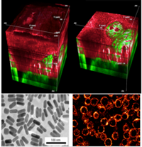
Top: 3D rendering of autofluorescence (red) and second harmonic generation (green) signal from human tongue tissue. Bottom Left: SEM image fo gold nanorods. Bottom Right: two-photon image of human cancer cells labeled with targeted gold nanorods.
|
Nonlinear microscopy is a potential technology for the diagnosis, screening, and monitoring of disease that allows real time, non-invasive imaging to be performed with subcellular resolution hundreds of micrometers deep in scattering tissues. Our lab, in collaboration with surgeons at the M.D. Anderson Cancer Center, has been investigating this technology as a means to classify oral tissue biopsies as normal, precancerous, or cancerous ex-vivo. Using two-photon and second-harmonic generation microscopy, we have generated 3D maps of endogenous fluorescence from the samples, which provide morphological and functional information for biopsy case-finding. To improve the sensitivity of this technique, we are introducing novel contrast agents, such as gold nanospheres and nanorods, which are specifically targeted to proteins which are overexpressed in cancerous tissues. Furthermore, the increased brightness from these exogenous contrast agents allow for endoscopic imaging of tissues in our less-sensitive, but more clinically friendly miniaturized nonlinear microscope. |
|